


 Islands")


")


")


")


")
")


 Germany
 Japan
Japan
 United Kingdom
 China
China
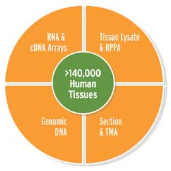
Comprehensive Human Cancer and Normal Tissue Products
OriGene has developed comprehensive human cancer and normal tissue products from its biorepository of over 140,000 high quality human tissues.
TissueScan™ Array Products
- TissueScan™ Tissue cDNA Array for differential gene expression profiling.
- Tissue Microarray (TMA) for multiplex histological analysis
TissueFocus™ Individual Products
- TissueFocus Control Slides - New product! We now have positive and negative control slides available for ALK, PD1 and PDL1 developed specifically for IHC, with more soon to be added.
- TissueFocus™ Search Tool allows you to search over 30,000 total RNA, genomic DNA, protein lysates, FFPE and frozen sections from human tissue samples. Case-matched tumor and normal samples are available
- Tissue total RNA and genomic DNA Samples were derived from our high quality frozen tissues after rigorous QC testing. Agilent Bioanalyzer 28S/18S ratio, Electropherogram, A260/A280 ratio and PCR images available upon request. Samples are offered in 5 ug aliquots.
- Tissue Protein Lysate Protein lysates were generated using a Modified RIPA buffer (no SDS) in the presence of protease and phosphatase inhibitors. Protein quantification is performed using the BCA Protein Assay. Samples are offered in 150 ug aliquots.
- Tissue Sections Frozen and FFPE tissue sections can be used for various applications such as IHC and ISH. Each section is freshly cut onto a SuperFrost positively charged glass slide, and offered as a set of 5 slides (each 5 micron thickness).
- TissueFocus™ Block available in formalin-fixed, paraffin embedded OR frozen, OCT-embedded formats
OriGene’s comprehensive tissue products provide researchers ready-to-use tools for their studies in biomarker discovery and validation, cancer drug target identification and validation, companion diagnostic assay development, and personalized medicine for the cancer patient. Tissues are banked under strict collection protocols and undergo rigorous quality control to ensure each source block’s unparalleled quality.
- Collected from major US institutions under strict IRB and ethical consenting practices
- Maintained in a monitored environment and bar-coded for tracking purposes
- Each tissue source block includes following clinical information
- Abstracted pathology report
- Tissue of origin / site of finding
- Disease staging
- Digital H&E image
- Donor’s basic demographic information
- Learn more about the quality control of human tissue products
Recent Citations using our Tissues
|
1. cfDNA methylome profiling for detection and subtyping of small cell lung cancers Published in Nature Cancer, 2022 Aug, using CD563113 |
PubMed: 35941262 |
|
2. Aggressive PDACs show hypomethylation of repetitive elements and the execution of an intrinsic IFN program linked to a ductal cell-of-origin Published in Cancer discovery, 2022 Jun, using CD564011 |
PubMed: 33060108 |
|
3. Abrogation of self-tolerance by misfolded self-antigens complexed with MHC class II molecules Published in Science Advances, 2022 Mar, using CS804207 |
PubMed: 35245125 |