Human Seprase/FAP ELISA Kit
USD 705.00
3 Weeks*
Size
Other products for "FAP"
Specifications
| Product Data | |
| Assay Type | Sandwich ELISA kit of Quantitative Detection for Human Seprase/FAP |
| Assay Length | 3.5 hours incubations; 1 hour washing and analyzing samples |
| Signal | Colorimetric |
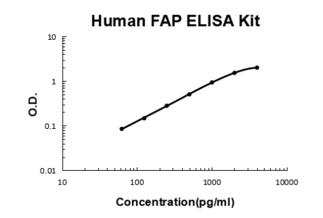
| Curve Range | 62.5pg/ml-4000pg/ml |
| Sample Type | Cell culture supernates, serum and plasma(heparin, EDTA). |
| Sample Volume | 100 ul |
| Specificity | This kit is used for quantitative detection of human Seprase |
| Sensitivity | <10pg/ml |
| Reactivities | Human |
| Cross Reactivity | There is no detectable cross-reactivity with other relevant proteins. |
| Interference | No significant interference observed with available related molecules. |
| Components |
|
| Background | FAP(Fibroblast Activation Protein, Alpha) also known as FAPA or SEPRASE, is an inducible cell surface glycoprotein that was originally identified in cultured fibroblasts using monoclonal antibody F19. The protein encoded by this gene is a homodimeric integral membrane gelatinase belonging to the serine protease family. The FAP gene is mapped on 2q24.2. FAP is most closely related to DPPIV and they share about 50% of their amino acids. FAP is catalytically active as a 170kD dimer and has dipeptidase and gelatinase activity. Its gelatinase activity requires a glycine in P2 position.FAP-alpha shows 48% amino acid identity with dipeptidyl peptidase IV and 30% identity with DPP4-related protein. Northern blot analysis detected a 2.8-kb FAP-alpha mRNA in fibroblasts. Depletion of FAP-expressing cells, which made up only 2% of all tumor cells in established Lewis lung carcinomas, caused rapid hypoxic necrosis of both cancer and stromal cells in immunogenic tumors by a process involving interferon-gamma and tumor necrosis factor-alpha. |
| Gene Symbol | FAP |
| Gene ID | 2191 |
Documents
| SDS |
{0} Product Review(s)
0 Product Review(s)
Submit review
Be the first one to submit a review
Product Citations
*Delivery time may vary from web posted schedule. Occasional delays may occur due to unforeseen
complexities in the preparation of your product. International customers may expect an additional 1-2 weeks
in shipping.


 Germany
 Japan
Japan
 United Kingdom
 China
China
