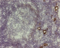
VAP1 (AOC3) Mouse Monoclonal Antibody [Clone ID: 174-5]
Specifications
| Product Data | |
| Clone Name | 174-5 |
| Applications | FN, IF, IHC |
| Recommended Dilution | Immunohistochemistry on frozen sections: Tissue sections were fixed in acetone before staining with 174-5. As positive control anti-VAP-1 mAb 2D10 was used and as negative control an isotype matched irrelevant mAb. (Ref.1). Typical starting working dilution is 1:50. Flow cytometry: Stains Ax cells stably transfected with VAP-1 cDNA.. As negative control mock transfected cells were used.(Ref.1). Typical starting working dilution is 1:50. Functional assay: Inhibits lymphocyte infiltration in liver allograft rejection. The antibody was administered intravenously at a concentration of 2 mg/kg. A irrelevant isotype-matched antibody served as a negative control. (Ref.1). Immunoflourescence (Ref1,2). Positive control: Ax cells stably transfected with VAP-1 cDNA. (Ref. 1). Negative control: Mock transfected Ax cells. (Ref. 1). |
| Reactivities | Human, Rat |
| Host | Mouse |
| Isotype | IgG1 |
| Clonality | Monoclonal |
| Immunogen | Purified vessels from human peripheral lymph nodes (ref 1) |
| Specificity | This antibody detects Vascular Adhesion Protein-1 (VAP-1). |
| Formulation | PBS State: Purified State: Liquid 0.2 µm filtered Ig fraction Stabilizer: 0.1% bovine serum albumin |
| Concentration | lot specific |
| Purification | Protein G |
| Conjugation | Unconjugated |
| Storage | Store at 2 - 8 °C. |
| Stability | Shelf life: one year from despatch. |
| Gene Name | amine oxidase, copper containing 3 |
| Database Link | |
| Background | Vascular Adhesion Protein-1 (VAP-1) is a glycosylated homodimeric membrane protein consisting of two 90 kDa subunits connected by disulfide bonds. It contains a short N-terminal cytoplasmic tail, a single membrane-spanning domain and a large extracellular part. A soluble form of VAP-1 (sVAP-1) has been described, which presumably results from the proteolytic cleavage of membrane-bound VAP-1. Structurally VAP-1 belongs to enzymes called semicarbamizide-sensitive amine oxidases, which contain copper as a cofactor. These enzymes deaminate primary amines in a reaction producing hydrogen peroxide, aldehyde, and ammonia. |
| Synonyms | HPAO |
| Reference Data | |
Documents
| Product Manuals |
| FAQs |
| SDS |
{0} Product Review(s)
Be the first one to submit a review


 Germany
 Japan
Japan
 United Kingdom
 China
China


